Condition Guide · Atlanta, GA

When a calcium deposit turns a tendon suddenly, sharply painful

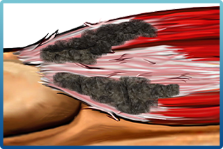
Calcific tendonitis is one of the more distinctive tendon problems: a deposit of calcium builds up inside a tendon — most often in the shoulder's rotator cuff — and can trigger pain that ranges from a dull background ache to an acute, hard-to-ignore flare. Understanding what the deposit is doing helps make sense of the treatment choices your specialist will walk you through.

6
Atlanta specialists
Image-
guided options
In-office
where appropriate

Patient Story

A Patient on the TenJet Experience

This patient describes their experience with the TenJet procedure, which specialists use across a range of chronic tendon conditions.

TenJet Patient Testimonial

Video courtesy of HydroCision, Inc., the maker of TenJet®. Individual results vary; this patient's experience is not a guarantee of outcome. Talk with a specialist about whether the procedure is appropriate for you.

Understanding The Condition

What the deposit is, and how it behaves

In calcific tendonitis, calcium crystals accumulate within a tendon. The deposit often passes through phases — forming quietly, then, in some people, breaking down in a way that provokes an intense but usually self-limiting flare. Because the pattern varies so much from person to person, imaging is central to sorting out what stage a deposit is in and what, if anything, needs to be done about it.

Most common in the shoulder, but it can occur in other tendons
Pain may be dull for months, then flare sharply for a stretch
Deposits are visible on X-ray and ultrasound, which guides treatment
Some deposits resolve on their own; others need intervention

Treatment Options

A range of approaches, matched to your case

Calcific tendonitis is treated in a stepwise way. Many people improve with activity modification, physical therapy, and anti-inflammatory measures. When symptoms persist, specialists use image-guided, in-office options — and the right one depends on the deposit, the tendon, and how much surrounding tendon has been affected. Your specialist will determine which procedure fits your situation.

Physical therapy and anti-inflammatory measures as a first line
Ultrasound-guided needle procedures aimed at the deposit itself
For associated tendon degeneration, a specialist may consider TenJet
The choice is individualized — imaging and a hands-on exam guide it

Where TenJet Fits

Addressing diseased tendon tissue directly

TenJet, made by HydroCision, Inc., is a percutaneous device that a specialist uses under ultrasound guidance to treat degenerated tendon tissue — the disorganized, weakened tissue that can accompany a long-standing tendon problem. Whether it is appropriate for a given case of calcific tendonitis is a decision only your specialist can make after examining you and reviewing your imaging.

Performed under real-time ultrasound guidance
A high-pressure saline stream emulsifies diseased tissue, sparing healthy fibers
Integrated suction removes that debris in the same pass
Entry is a small incision under local anesthesia — typically an in-office procedure
Reimbursed by Medicare and most private insurers when indicated
Read the technical details at hydrocision.com →

See The Procedure

TenJet® in Action

A general demonstration from HydroCision of how the TenJet procedure works, plus a look at how healthy tendon tissue differs from tissue that has degenerated over time.

TenJet Patient Animation — How the Procedure Works

Healthy tendon fiber structure diagram
Healthy Tendon
Tendinitis inflamed tendon diagram
Tendinitis
Tendinosis degenerated tendon fiber diagram
Tendinosis (Chronic Degeneration)

Diagrams and video courtesy of HydroCision, Inc.

Common Questions

Frequently Asked Questions

What is calcific tendonitis?

Calcium deposits forming within a tendon, often alongside degeneration. Deposits can be identified on ultrasound or radiographs and may cause considerable pain relative to their size.

Can the deposits be removed?

Published cases describe calcium deposits being broken up and extracted with the pressurised saline jet, with follow-up imaging showing resolution of the calcific debris.

Which tendons are commonly affected?

The rotator cuff most often, but published cases also document calcific tendinosis in the Achilles and elsewhere. Your physician determines the approach from imaging.

Is this the same as barbotage?

No. Barbotage uses needling and lavage to break up a deposit. This is a tenotomy procedure that removes diseased tissue, and your specialist decides which is appropriate.

This information is general and is not medical advice. Whether any procedure is appropriate for you can only be determined by a physician who has examined you and reviewed your imaging.

Speak Directly With Each Physician's Office
Call the office below that matches the specialist you'd like to see about calcific tendon pain.
CC
Sports Medicine & PM&R
Northside Hospital Orthopedic Institute
(770) 795-5620
MS
Non-Operative Orthopedics
Northside Hospital Orthopedic Institute
(770) 758-5059
KK
Dr. Kamal Kabakibou, MD
Interventional Pain Medicine
Center for Pain Management, Atlanta
(404) 882-9778
EO
Interventional Pain Medicine
OlympusMD Pain & Wellness
(770) 629-9023
OH
Interventional Pain Medicine
OlympusMD Pain & Wellness
(770) 629-9023
UN
PM&R & Spine
Resurgens Orthopaedics, Covington & Decatur
(404) 856-3706